Arthroscopy
Common Disorders we treat
- ACL Injury
- Meniscus Injuries
- PCL Injury
- Multi ligament injury
- Articular Cartilage lesions
- Patellar Instability
Services we offer
- ACL reconstruction
- Revision ACL reconstruction
- Meniscus surgery
- PCL Reconstruction
- Multi-ligament Reconstruction
- Cartilage restoration
- Patella (MPFL) Realignment
- Tibial/ Femoral Osteotomy
- Arthroscopic Loose Body Removal
ACL Injury
Anterior Cruciate Ligament
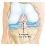
ACL is an important soft tissue structure inside the knee joint. Its function is to stabilise the knee joint by connecting between thigh bone (femur) and leg bone (tibia).ACL is frequently torn by twisting injury to the knee during sports activity and by being hit onto knee from falls.A completely torn ACL cannot heal on its own. Complete ACL tears are treated through key-hole surgery (Arthroscopy) via small incisions using a combination of fibre optics and small instruments.Partial ACL tears can heal with time. However, some patients with partial ACL tears may still have instability symptoms and needs surgical treatment.
Meniscus Injuries
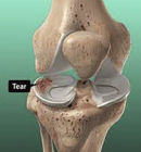
Meniscus is a pad of fibrocartilage that acts as a cushion within the knee joint. They serve as shock absorbers between the ends of the bones to protect the surface of the articular cartilage. Without a functioning meniscus, the articular cartilage is exposed to increased pressure and tends to wear earlier, leading to osteoarthritis.Meniscus injury is common, especially among people who play sports. A sudden twist, turn or collision can tear a meniscus. The Main symptom of this condition is the Knee pain, especially at the
side of the knee joint. Acute injuries are managed with Rest, Ice application, medications and physical therapy. If these treatments don’t work or if the injury is severe, surgery might be necessary.
PCL Injury
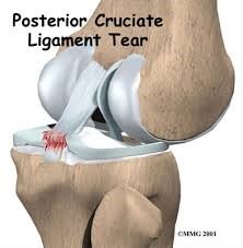
The posterior cruciate ligament (PCL) is located near the back of the knee joint. The PCL is the primary stabilizer of the knee and the main controller of how far backward the tibia moves under the femur. If the tibia moves too far back, the PCL can rupture.
The most common way for the PCL alone to be injured is from a direct blow to the front of the knee while the knee is bent.
The symptoms following a tear of the PCL can vary. Some patients may have a feeling of insecurity and giving way of the knee, especially when trying to change direction on the knee.
Less severe PCL tears are usually treated with a progressive rehabilitation program. If the PCL alone is injured, nonsurgical treatment may be all that is necessary. When other structures in the knee are injured, patients generally do better having surgery within a few weeks after the injury.
Multi ligament injury
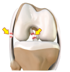
The 4 ligaments in and around the knee which are the main reasons for the stability are ACL, PCL, MCL and LCL. Normal tension within the ligament prevents abnormal movements within the knee joint. When two or more major ligaments of the knee are injured, it is termed as multi-ligamentous injury.
Such type of injury occurs as result of high velocity trauma like Road Traffic Accidents. Treatment for multi-ligament injury is always surgical reconstruction.
Articular Cartilage lesions
An articular cartilage injury is damage to the cartilage that lines the surface of joints (articular cartilage). The cartilage is smooth, white tissue that covers the ends of bones where they meet at a joint. This cartilage allows smooth movement of the joint. Injuries to joint cartilage can cause pain and decrease range of movement.
Treatment depends on the severity of the injury and the joint that is involved. Treatment options include: Drilling holes through the cartilage into the bone underneath it to improve blood flow, removing torn or damaged cartilage, replacing cartilage with a type of cartilage graft, replacing the entire joint.
Patellar Instability
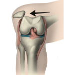
The kneecap (patella) is located in a groove in front of the lower end of the thighbone (femur). A patellar dislocation occurs when your patella slips all the way out of the groove.
The patellar dislocation results in the rupture of a ligament on the inner side of the knee called the MPFL(medial patellofemoral ligament) that keeps the kneecap in center of knee. Deficient MPFL can lead to recurrence of the dislocation even with trivial trauma. Less common factors such as altered limb alignment, flattening of the grove underlying the patella(trochlea dysplasia), Patella alta (relatively higher position of the patella) can also cause recurrence.
In general, the initial treatment of most patellar dislocations is non-operative with a dedicated rehabilitation program.
In those patients who have recurrent dislocations, the recommended treatment is surgery to stabilise the knee to the center of knee.
Services we offer
- ACL reconstruction
- Revision ACL reconstruction
- Meniscus surgery
- PCL Reconstruction
- Multi-ligament Reconstruction
- Cartilage restoration
- Patella (MPFL) Realignment
- Tibial/ Femoral Osteotomy
- Arthroscopic Loose Body Removal
ACL reconstruction
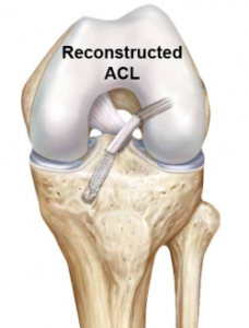
It is performed through key-holes (Arthroscopy) via small incisions using a combination of fibre optics and small instruments. In ACL reconstruction surgery, the torn ligament is replaced with a tissue graft, which will mimic the natural ACL. A portion of patient’s own tendon (patellar, hamstring, or quadriceps) is used to make a new ACL. A tunnel is drilled into both the tibia and femur. The graft is threaded across the knee and secured in place by using screws, buttons, staples or sutures. This restores the stability to the knee joint.
Revision ACL reconstruction
A revision ACL reconstruction is a second surgery needed to replace the deficient ACL graft. ACL revision surgery is a more challenging operation than a primary reconstruction.
Revision ACL reconstruction surgery is recommended when there is recurrent instability feeling of the knee, even after focussed rehabilitation and physiotherapy. Some patients may require a two
stage revision surgery, in which Bone grafting is done during the first surgery to fill up the previous surgery tunnels in the bones. New ACL ligament is reconstructed using one of the options: the
patellar tendon, quadriceps tendon, hamstring tendon, peroneus longus tendon or tendon from the opposite side knee.
Meniscus surgery
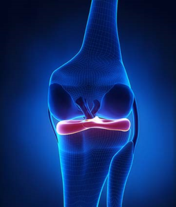
Meniscus is operated arthroscopically using small incisions (key-hole surgery) using an Arthroscope, which is a thin tube with light and video camera at its end.
Two types of Meniscus Surgery:
- Meniscus Repair – The Torn meniscus portions are stitched together using special instruments and meniscus is allowed to heal.
- Partial Meniscectomy – The damaged meniscal tissue is trimmed and removed leaving the healthy meniscus in place.
Recovery after the Meniscus Surgery
- The recovery time depends on the type of surgery performed.
- Full recovery from Meniscus repair takes between six weeks to twelve weeks.
- After Partial Meniscectomy, the recovery is much quicker.
PCL Reconstruction
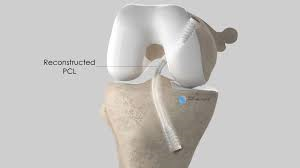
In this Surgery, torn PCL ligament is replaced using a piece of tendon or ligament (autograft). The surgery is performed through key hole incisions (Arthroscopy).
Multi-ligament Reconstruction
Multi-ligament injury always require surgery in the form of arthroscopic and open ligament reconstruction.
The reconstruction requires autografts (patient’s own) harvested from the same or opposite knee.
Generally a waiting period of 10-14 days is required for the soft tissue swelling to subside before surgery. Surgery can be done in single stage or in two stages, depending upon the severity of the injury.
Cartilage Restoration
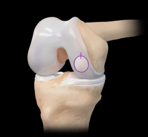
Defects smaller than 2 cm have the best prognosis. Treatment of such defects include arthroscopic surgery involving drilling and causing microfractures(creating small holes). This procedure stimulates healing response by marrow cells that produce cartilage at the area of the defect almost similar to the native cartilage.
For slightly larger defects, it may be necessary to transplant cartilage from other areas of the knee called mosaicplasty/OATS- Osteochondral autograft transfer.
For very large defects, it is necessary to harvest the patients cartilage cells in the 1st stage operation which are then cultured followed by implantation of these cells about 3 weeks later. This procedure is called autologous chondrocyte implantation(ACI).
Patella (MPFL) Realignment
Patellar instability surgery involves: Reconstruction of the MPFL ligament using autograft. This is done arthroscopically using key-hole incisions.
Release of tight tissues on the outer side of the knee.
Moving the area where your patellar tendon attaches to your shin bone (tibial tubercle transfer).
Tibial Osteotomy
The procedure involves a cut through the upper part of the tibial bone (Osteotomy). After the bone is cut, the two sides of the tibia are separated to form a wedge-shaped opening. This opening is then filled with bone graft and the bone is stabilised using special plates. This procedure changes the overall axis of the lower leg and shifts the load onto the healthier side within the knee joint.

